Resident quick read
- Name the abnormal component: MTSL, SPL, ISL, central plantar plate or collateral ligament—not a generic “plantar plate tear.”
- Edema with continuous fibers is a sprain. A partial tear needs a demonstrable focal fiber defect or convincing attenuation with a fluid cleft.
- Sesamoid marrow edema is nonspecific: stress reaction, fracture, bipartite synchondrosis injury, osteonecrosis, arthrosis, gout and infection can overlap.
- A smooth, well-defined distal plantar plate recess is normal and should not be called a tear.
- Static MRI demonstrates structural injury; it does not independently prove dynamic instability.
Scope and method
This is a narrative educational review for radiologists and trainees interpreting MRI of the hallux sesamoid complex and first metatarsophalangeal (MTP) joint. It emphasizes applied anatomy, direct and indirect imaging signs, component-level injury localization and clinically useful reporting language. It is not a systematic review and does not replace clinical assessment.
The main anatomical framework is drawn from Hallinan et al. and the high-resolution MRI–histology study by Wang et al. Clinical examples are individual open-access case reports and should be treated as illustrations, not universal diagnostic criteria.
1. Applied anatomy: think of a complex, not two bones
The medial (tibial) and lateral (fibular) sesamoids are embedded in the medial and lateral heads of the flexor hallucis brevis (FHB), beneath the first metatarsal head. Each articulates with a corresponding plantar facet of the metatarsal head. The flexor hallucis longus (FHL) courses between the sesamoids and continues to the plantar base of the distal phalanx. The sesamoids bear load and improve FHB mechanical leverage.1
The plantar stabilizing apparatus includes the sesamoids, fibrocartilaginous plantar pad, central plantar plate, intersesamoid ligament (ISL), paired metatarsosesamoid ligaments (MTSLs), paired sesamoid-phalangeal ligaments (SPLs), plantar capsule and supporting musculotendinous structures.

Original schematic · plantar view
MTSLs attach proximally at the sesamoids; SPLs attach distally toward the plantar proximal phalanx. Simplified and not to scale.
1.1 Terminology: how to use “plantar plate”
The first MTP plantar plate is not a simple equivalent of the lesser-MTP plantar plate. Hallinan et al. describe a fibrocartilaginous pad investing the sesamoids and related to the plantar capsule. Wang et al., using high-resolution 3T MRI with histology, describe four components: central plantar plate, ISL, SPLs and MTSLs.1, 2
Adjacent collagen fibers may interdigitate or merge at attachments. Name the component that is visibly abnormal when image quality permits. If the components cannot be separated, describe the dominant region and state the limitation rather than inventing an exact tear location.

1.2 Proximal and distal attachments
- MTSLs: metatarsal neck/head region to proximal margins of the sesamoids.
- SPLs: distal sesamoids to plantar base of the proximal phalanx.
- ISL: spans between the sesamoids and is continuous with the central plantar plate in the midsagittal anatomical description.
- Collateral ligaments: main medial and lateral bands from metatarsal head to proximal phalangeal base; distinct from accessory sesamoid ligaments.

1.3 Supporting tendons and muscles
The medial and lateral FHB heads attach to their respective sesamoids. Abductor hallucis reinforces the medial complex and joins the medial FHB region; adductor hallucis supports the lateral complex and joins the lateral FHB region. The main collateral ligaments and accessory sesamoid ligaments are separate structures and should not be conflated.
2. MRI technique and plane-specific assessment
Use a small field of view centered on the first MTP joint, thin sections and high-resolution fluid-sensitive imaging in three planes. Include at least one T1-weighted sequence for marrow architecture, sclerosis and fracture-line assessment.
| Sequence | Primary reporting purpose |
|---|---|
| T1-weighted | Marrow replacement, fracture line, sclerosis and chronic structural change. |
| PD/intermediate-weighted FS | Ligaments, tendons, marrow edema, capsule and cartilage-adjacent abnormalities. |
| T2 FS / STIR | Edema, fluid, synovitis and bursal distention. |
| Dedicated oblique/parasagittal | MTSL and SPL attachments when routine planes do not show them adequately. |
Consider weight-bearing radiographs, contralateral comparison or dorsiflexion stress views when bipartite sesamoid, traumatic diastasis or dynamic instability remains uncertain. CT clarifies cortical fracture morphology and sclerosis; ultrasound can assess superficial structures dynamically.
Hallinan et al. use “coronal” for the short-axis plane through the great toe and “axial” for the long-axis plane. Local scanner labels may differ. Confirm true anatomic orientation rather than relying on the sequence label.
SHORT-AXIS / CORONAL
- MTSLs and sesamoid attachments
- Intersesamoid ligament
- Central plantar plate and paired SPLs
- Sesamoid alignment and FHB heads
SAGITTAL / PARASAGITTAL
- SPLs to proximal phalangeal base
- Central plantar plate profile
- FHL course
- Sesamoid-metatarsal articulation
LONG-AXIS / AXIAL
- Medial and lateral collateral ligaments
- Abductor and adductor hallucis
- FHB heads and transverse relationships
- Dorsal extensor mechanism


3. Normal MRI atlas: cadaveric anatomy

4. Osseous sesamoid disease
4.1 Stress reaction and sesamoiditis
Stress-related change typically shows fluid-sensitive marrow hyperintensity, variable T1 hypointensity and adjacent soft-tissue edema without a discrete fracture line or cortical interruption. “Sesamoiditis” is etiologically nonspecific. Prefer “stress-related marrow edema” when repetitive loading is plausible but no cause is established. Marrow edema must be interpreted with morphology, articular surfaces and surrounding soft tissues.
4.2 Fracture versus bipartite sesamoid
| Feature | Bipartite sesamoid | Acute fracture |
|---|---|---|
| Margins | Smooth, rounded, corticated | Irregular, often noncorticated |
| Interface | Synchondrosis | Fracture cleft |
| Marrow edema | Usually absent unless symptomatic or injured | Common |
| Contralateral foot | May show similar variant | Usually no matching fracture |
Neither smooth margins nor bilaterality excludes symptomatic synchondrosis injury or fracture through a pre-existing variant. If MRI cannot separate acute diastasis from fracture, say “indeterminate” and suggest prior imaging, contralateral radiographs or CT.
4.3 Osteonecrosis
Look for sclerosis, altered trabecular architecture, fragmentation, flattening/collapse and secondary articular degeneration. Early osteonecrosis overlaps with stress reaction; marrow edema or T1 hypointensity alone is insufficient. Radiographs or CT can characterize sclerosis and cortical architecture.

4.4 Sesamoid-metatarsal arthrosis and first MTP osteoarthritis
Assess medial and lateral sesamoid-metatarsal joints independently. Look for cartilage thinning/loss, subchondral edema and cysts, sclerosis, osteophytes and surface irregularity. Do not conflate this with osteoarthritis of the principal first MTP articulation (hallux rigidus), which may show joint-space and cartilage loss, dorsal/marginal osteophytes and subchondral cysts.
5. Plantar capsuloligamentous injury and turf toe
Turf toe is a hyperextension injury of the plantar stabilizing complex of the first MTP joint. SPLs are particularly vulnerable. MRI should describe the structural injury present rather than use “turf toe” as a substitute for anatomic localization.4, 7
5.1 Structural severity framework
Describe the structural severity spectrum, but do not equate it automatically with a formal clinical turf-toe grade or functional instability.
Preserved
Normal low-signal fibers; no edema or discontinuity.
Sprain
Thickening or edema with preserved fiber continuity.
Partial tear
Focal fiber defect or convincing attenuation with a fluid cleft.
Complete tear
Full-thickness discontinuity; assess retraction, avulsion and sesamoid migration.
- Sprain: periligamentous edema and thickening with preserved fiber continuity.
- Partial tear: focal fiber discontinuity, or convincing focal attenuation/contour loss with a fluid-signal cleft in the expected ligament course.
- Complete tear: full-thickness discontinuity; assess retraction, avulsion, sesamoid fracture, diastasis and proximal migration.
MRI demonstrates structural disruption. Clinical examination, stress radiographs or dynamic ultrasound assess instability. Do not infer prognosis, treatment indication or return-to-sport timing from an MRI pattern alone.
5.2 Component-by-component review
- MTSL: assess medial and lateral separately. A focal fluid cleft at the sesamoid attachment supports partial proximal disruption.
- SPL: trace each side in sagittal/parasagittal images. Document discontinuity, attachment, retraction, avulsion and sesamoid position.
- ISL: assess on short-axis images for thickening, edema, partial/full discontinuity and sesamoid diastasis.
- Central plantar plate: assess metatarsal neck origin and distal phalangeal attachment. Focal discontinuity is more specific than signal change alone. A smooth distal recess is normal.
- Collateral ligaments: assess medial and lateral main bands separately from accessory sesamoid ligaments.
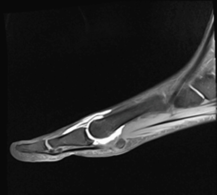
6. Collaterals, tendons, muscle attachments and extrinsic causes
6.1 Tendons and muscle
- FHB: assess medial and lateral heads separately, including sesamoid attachments and distal continuation.
- FHL: trace between the sesamoids and distally. Trace sheath fluid can be physiologic; tenosynovitis requires disproportionate fluid, synovial thickening or tendon abnormality.
- Abductor/adductor hallucis: insertional edema without discontinuity is enthesopathic/insertional change, not a tear.
- EHL/EHB: assess tendinosis, tear, tenosynovitis, lateralization and dorsal attrition.
6.2 Hallux valgus and hallux rigidus
Assess hallux alignment, sesamoid subluxation/rotation, medial capsular attenuation, collateral integrity, extensor tendon lateralization and secondary cartilage/plantar complex attrition. Lateral sesamoid displacement may reflect altered first-ray alignment rather than intrinsic sesamoid disease.
6.3 Gout, inflammatory arthropathy and infection
MRI is sensitive for marrow, synovial and soft-tissue involvement but not specific for urate or infection in isolation. Look for erosions, synovitis, tophaceous deposits and marrow edema in inflammatory arthropathy. Infection becomes more concerning with confluent T1 marrow replacement, cortical destruction, synovitis, abscess, sinus tract or tendon-sheath involvement. Correlate with clinical, laboratory, radiographic and aspiration findings as appropriate.
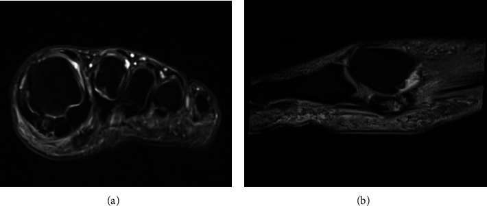
6.4 Bursitis and digital nerve findings
Distinguish intermetatarsal bursitis from adventitial plantar bursitis, joint recess fluid and FHL sheath fluid. A Morton neuroma requires a focal nerve-related mass or enlargement; perineural fat stranding alone is nonspecific. Specify the correct nerve and webspace, particularly the common plantar digital nerve in the first webspace.
7. Clinical MRI case examples
These are individual published cases reproduced under their stated open-access licences. They illustrate imaging patterns and are not diagnostic criteria by themselves.
Osteonecrosis: fluid-sensitive hyperintensity and corresponding T1 hypointensity may accompany chronic structural change. Sclerosis, fragmentation and collapse on radiographs/CT strengthen the diagnosis.
Bipartite sesamoid with edema: marrow and periarticular edema can occur in a bipartite sesamoid; the underlying cause may be stress-related, inflammatory or articular.
Turf toe: severe plantar capsuloligamentous disruption should prompt structure-by-structure assessment, including sesamoid position and associated avulsion/fracture.
8. Differential diagnosis at a glance
| MRI pattern | Main differential | Discriminator |
|---|---|---|
| Sesamoid marrow edema | Stress reaction, fracture, osteonecrosis, arthropathy, infection | Fracture line, sclerosis/architecture, articular pattern, cortical destruction/collection |
| Fragmented sesamoid | Bipartite variant, fracture, traumatic diastasis, osteonecrosis | Cortication, margin morphology, edema, sclerosis and collapse |
| Plantar soft-tissue signal | Ligament injury, FHB injury, bursitis, neuroma, infection | Expected anatomic course, fiber continuity, focal mass, bursal location, collection |
| Subchondral metatarsal head edema | Reactive osteoarthritis, stress injury, osteochondral lesion | Cartilage defect, fracture line, subchondral cyst/osteophyte pattern |
9. Pitfalls and systematic checklist
- Do not call a partial tear from edema alone; look for focal fiber discontinuity or a convincing fluid cleft.
- Do not use “plantar plate tear” as a substitute for localizing MTSL, SPL, ISL or central plantar plate.
- Do not mistake the smooth distal plantar plate recess for a tear.
- Do not call a bipartite sesamoid a fracture without evaluating cortication, interface morphology and edema.
- Do not diagnose osteonecrosis from marrow edema or T1 hypointensity alone.
- Evaluate the main first MTP cartilage and both sesamoid-metatarsal articulations separately.
- Distinguish main collateral ligaments from accessory sesamoid ligaments.
- Do not overcall trace FHL sheath fluid as tenosynovitis.
- Perineural fat stranding without focal nerve enlargement is not a definite Morton neuroma.
- Static MRI cannot prove dynamic instability.
Systematic review checklist
- Alignment: hallux valgus/varus, MTP subluxation, sesamoid position/rotation/retraction.
- Sesamoids: morphology, fracture, edema, sclerosis, fragmentation and displacement.
- Articulations: main MTP and medial/lateral sesamoid-metatarsal cartilage.
- Plantar complex: MTSL, SPL, ISL, central plate and capsule.
- Collaterals: medial/lateral main collateral and accessory ligaments.
- Tendons/muscles: FHB heads, FHL, abductor/adductor hallucis, EHL/EHB.
- Soft tissues: bursitis, synovitis, nerve, collection and susceptibility artifact.
- Associated injury: metatarsal head, proximal phalanx base, avulsion and osteochondral fragment.
10. Report library and impression order
Use these as starting points. Adapt to the exact structure, side, attachment and certainty visible on the study.
Low-grade ligament sprain
Mild thickening and intrasubstance fluid-sensitive signal of the [medial/lateral] [MTSL/SPL/collateral ligament], with preserved fiber continuity, consistent with a low-grade sprain.
Partial-thickness tear
Focal partial fiber discontinuity and surrounding edema of the [medial/lateral] [MTSL/SPL] at its [metatarsal/sesamoid/phalangeal] attachment, compatible with a partial-thickness tear. No retraction.
Sesamoid stress reaction
Marrow edema within the [medial/lateral] sesamoid without a discrete fracture line or cortical disruption, compatible with stress-related change.
Sesamoid fracture
[Acute/subacute] [nondisplaced/displaced] fracture of the [medial/lateral] sesamoid with surrounding marrow and periosseous edema.
Sesamoid-metatarsal osteoarthritis
[Mild/moderate/advanced] [medial/lateral] sesamoid-metatarsal osteoarthritis with [cartilage thinning/loss], subchondral [edema/cysts/sclerosis] and marginal osteophytes.
Digital nerve abnormality without definite neuroma
Mild perineural fat stranding surrounding the common plantar digital nerve in the [first/second] intermetatarsal webspace, without focal nerve enlargement or a discrete neuroma.
Insertional enthesopathic change
Mild insertional enthesopathic edema involving the [medial/lateral] [FHB/abductor hallucis/adductor hallucis] attachment, without tendon discontinuity.
Impression order
- Dominant structural injury: exact ligament, side, attachment and completeness.
- Osseous injury: sesamoid/metatarsal site, fracture, displacement or structural osteonecrosis.
- Articular degeneration: distinguish main first MTP from sesamoid-metatarsal arthrosis.
- Associated tendon, muscle, nerve, bursal or soft-tissue abnormality and meaningful artifact limitation.
11. Evidence table: what is supported and what needs correlation
| Statement | Evidence basis | Limitation |
|---|---|---|
| Four plantar components can be localized on high-resolution MRI | MRI-histology correlation and anatomy studies 1, 2 | Routine thick-slice or edematous studies may limit separation. |
| SPLs are commonly injured in turf toe | Imaging reviews 4, 7 | Clinical grade requires examination. |
| Edema with continuous fibers is sprain, not tear | Structural MRI reporting principle; evaluate direct fiber signs. | Partial tears may be difficult to resolve at routine spatial resolution. |
| Fracture versus bipartite diastasis may overlap | Imaging reviews and case literature 1, 3 | Prior imaging, contralateral radiographs or CT may be needed. |
| Osteonecrosis requires structural support | Case-level and review evidence 5 | Radiograph/CT correlation helps show sclerosis and fragmentation. |
| MRI independently proves gout, infection or dynamic instability | Not supported. | Clinical, laboratory, aspiration, stress radiograph or dynamic ultrasound correlation. |
12. Limitations
- Much of the detailed anatomical correlation comes from cadaveric or high-resolution MRI studies; routine clinical MRI may not reproduce that spatial resolution.
- Uniformly validated MRI criteria for partial injury of individual first-MTP plantar structures remain limited.
- MRI cannot independently determine dynamic instability, pain generator, treatment indication or athletic prognosis.
- This is a narrative review, not a systematic review; clinical images are individual illustrative cases.
Self-test: five reporting questions
1. Thickening and edema of the medial SPL, with continuous fibers: what is the best description?
Low-grade sprain. A partial tear needs focal fiber discontinuity or convincing focal attenuation with a fluid cleft.
2. A smooth, well-defined distal recess at the plantar plate insertion is what?
A normal anatomical recess; do not call it a tear.
3. Marrow edema and low T1 signal without sclerosis or collapse: can osteonecrosis be diagnosed confidently?
No. Signal alone is insufficient; stress reaction and early osteonecrosis overlap.
4. Can MRI always distinguish bipartite synchondrosis injury from fracture?
No. Use prior imaging, contralateral radiographs or CT when morphology remains indeterminate.
5. Can static MRI establish dynamic instability?
No. Describe structural disruption; use examination, stress radiographs or dynamic ultrasound for instability.
References and image rights
- Hallinan JTPD, Statum SM, Huang BK, et al. High-resolution MRI of the first metatarsophalangeal joint: gross anatomy and injury characterization. RadioGraphics. 2020;40(4):1107–1124. DOI · Article
- Wang JE, Bai RJ, Zhan HL, et al. High-resolution 3T MRI and histological analysis of the capsuloligamentous complex of the first MTP joint. J Orthop Surg Res. 2021;16:638. DOI · Article
- Lombard C, Gillet R, Rauch A, et al. Hallux sesamoid complex imaging: a practical diagnostic approach. Skeletal Radiol. 2020;49(12):1889–1901. DOI · Article
- Claikens T, Rezaie W, Simons P. Turf toe injury: a pictorial review and a didactic case report. Semin Musculoskelet Radiol. 2024;28(2):213–217. DOI · Article
- Saucedo JG, Pai R, Gerstner JB. An overlooked cause of forefoot pain in athletes: avascular necrosis of the lateral hallucial sesamoid. Radiol Case Rep. 2025. DOI · Article
- Beraldo DO, et al. Bilateral sesamoiditis as first manifestation of gout. Case Rep Orthop. 2020;2020:8890549. DOI · Article
- Limaye N, Sethi M, Ayyaswamy B. Outcomes of surgical management of turf toe: 12-year results. Cureus. 2024;16(4):e57808. DOI · Article
Further reading
- Srinivasan R. The hallucal-sesamoid complex: normal anatomy, imaging, and pathology. Semin Musculoskelet Radiol. 2016;20(2):224–232. DOI · Record
- Theumann NH, Pfirrmann CWA, Mohana Borges AVR, et al. Metatarsophalangeal joint of the great toe: normal MR, MR arthrographic, and MR bursographic findings in cadavers. J Comput Assist Tomogr. 2002;26(5):829–838. DOI · Record
- Sanders TG, Rathur SK. Imaging of painful conditions of the hallucal sesamoid complex and plantar capsular structures of the first metatarsophalangeal joint. Radiol Clin North Am. 2008;46(6):1079–1092. DOI · Record
Evidence-access note: Hallinan et al. and Wang et al. were reviewed in full text. Lombard et al. was bibliographically verified and its accessible metadata/abstract reviewed; complete publisher text was not available during preparation. Srinivasan, Theumann and Sanders are listed as further reading, not as full-text-extracted sources. A subspecialist author should perform final claim-by-claim review before external scientific endorsement.
United TeleRadiology · Clinical Masterclass Series
Clinical education for better imaging and reporting.
Explore the United TeleRadiology Blogs library or contact our team about clinical education and professional collaboration.